To View Part 1 or 5 First
Click on Links Below
Malassezia: What The Photos Reveal – (PT 1)
Malassezia: What The Photos Reveal – (PT 5)
*
* UNDER THE MICROSCOPE *
*
* Malassezia: One -Two or Many *
* What The Photos Reveal – PT 6 *
*
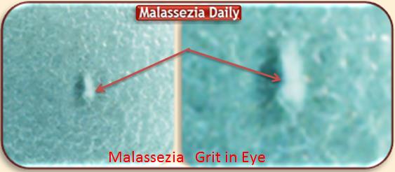
* Malassezia: Grit in the Eye *
What Photos Reveal Under Microscope
*
PT 1 Grit in the Eye
Normally the gritty particles in the eye – a plethora of them!
are wrapped and protected within the Liquid Biofilm and are not visible.
They can be isolated and become visible
by rubbing between fingers until liquid is dried.
This one was sitting in the liquid, right on the retina of my eye
and must have been rather mature
because when i pulled it out and rubbed it it was easier to isolate
and more visually distinguishable than usually
so i rushed it to my … Laboratory for closer Microscope examination
or is it in legal terms… ‘Cross Examination’ ? Lol!
‘Ve got Vays to make vyou Talk!’ 🙂
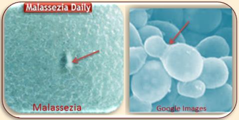
Grit in the Eye Viewed under Digital Microscope
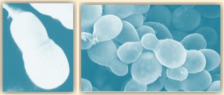
Below Google Official Lab Photos for Comparison of Shape
Obviously Practically Identical despite poor photo Focussing
The arrows indicating respective places of Cloning Collarets
Malassezia Eye Grit Under Biological Microscope
The Same Gritty Particle viewed under Biological Microscope.
I know the view under each Microscope is usually widely different
but what i saw surprised me in Three distinct ways!
One: I did not quite expect to see something so entirely different
Two: Hyphae … so quite Developed!…
Three: Looking Identical to the Red Hyphae photo
i had published in much earlier Entries a couple of years back!
I Suppose that’s the idea of Cloning!…
(The Original Hypha Photo i Posted way Back)
(PART 7: * Malassezia: Gritty Liquid in the Eye * )
==============================================
Recommended Reading Entries in Order of Posting
* MALASSEZIA BLOGS *
* Malassezia Blog *
Photos – History of Treatments and Results
* Malassezia Daily *
New Photos – New Discoveries
Treatments and Results
Malassezia Issues – Related Topics
Readers Questions Answered
Malassezia and Medical Research
Health and Immunity – Nutrition Diet
=============================================


