To View Part 1 or 7 First
Click on Links Below
Malassezia: What The Photos Reveal – (PT 1)
Malassezia: What The Photos Reveal – (PT7)
* * *
* UNDER THE MICROSCOPE *
*


* Malassezia: One -Two or Many *
* What The Photos Reveal – PT 8 *
*
Malassezia
* Vaginal and Faecal – Intestinal Samples *
*
Malassezia in Vaginal Sample
At this stage i am unable or rather Unwilling
to produce current Photos of either Vaginal or Faecal Samples
Because:
a) I have already published some earlier stage photos from Both areas
in the Photos section in the *Malassezia Blog*
b) I Know with Absolute Certainty by the Traffic and Symptoms
what exactly exists almost perpetually in Both areas, and
c) It would Require me to leave them Unprotected for some time
and allow Malassezia to take hold and be tortured during that time.
After the ‘Malassezia – Acidophilus Round 2 & The 8 Hour Kill?’
I Am Not willing to go by design through any similar experience
I do not feel that any sacrifice of going backwards a few steps
with Resulting Consequences, are worth the Pain and Inconvenience
just for producing photographic demonstration.
For Readers Benefit though i have Re-visited, Zoomed
and worked with the Original Photos of Both areas
-photographed back then with a very poor quality camera-
in order to bring forth the Common Elements.
Despite the Original poor Quality of Photos
it is still ALL there in Plain Clear View
and all i have to do is present the Original Entry Photos
with Additional Details and Comments in this section
and then we will All be Happy -Lol!
without any Distress caused to the Poor Ol ‘Damsel’ 🙂
Vaginal Sample
(Click On Photos to Enlarge)

A: It looks very much like the Orange sticky liquid Biofilm
in which the Solidifying Grit or Fatty Blobs are usually wrapped
and which acts as the Carrying and Protecting ‘Amniotic Fluid’.
B: Long Hyphae extending from (C)
C: A mature unit with Unfolding Hyphae
D: Insert of an earlier published of similar one for shape comparison
Below, on Top part of photo a smaller size visible, with unfolding Hyphae

Below Zoomed makes Visible the Beaded Cloning inside the Hyphae

Faecal Sample 1
Maroon Arrows point to the Orange Sticky ‘Amniotic’ Biofilm Liquid
Green Arrows point to Long Hyphae

(Click on Photos to Enlarge)
Circular Beaded Eyelet
Below Zoomed makes Visible one Circular Beaded Eyelet

And if i see it correctly at the edge of the Fluid a Beaded Hyphen
Faecal Sample 2

Top Part of Photo Below
Green Arrows: Long Hyphae
Purple Arrow: A Beaded Hyphen
Orange C Shapes: Malassezia Orange / Greenie Sticky Biofilm Liquid

(Click on Photos to Enlarge)
Bottom Part of Photo
Green Circular Shapes point to a plentiful of Spaghetti Hyphae
Purple Arrows point to Extra Long -Well …‘Fed’
and Well Developed- Hyphae

(Click on Photos to Enlarge)
Abundant indication of what i have been saying all along throughout my Entries:
a) There are a Lot of Rich Nutrients for Malassezia in that area
b) It Gathers there in Concentrated quantities causing Congestion
Constipation, Blocking, Anal Itch and Leg jerking
and that’s why Enemas are Helpful
in Bringing Relief to the Congestion, Restless Leg
and Getting Rid of a Great Bulk of Presence
with just a…‘Flash’ or Two! Lol! 🙂
Considering the photo above shows such
a Compact Presence of ‘Spaghetti’ Hyphae,
in a sample smaller than a Pea sized pooh
smeared thin on a slide
one can imagine how much would be contained
in one ‘Regular size’ motion which
-compared to how much even a small 2 Ltr Enema would Flash out-
is only the Tip of the Iceberg!
* * *
Below Malassezia Lab Images
included for Hyphae Shape and Beaded Appearance comparison
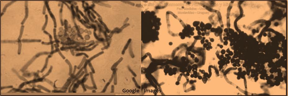
( PART 9: *The Big Surprise! * )
==============================================
Recommended Reading Entries in Order of Posting
* MALASSEZIA BLOGS *
![]()
* Malassezia Blog *
Photos – History of Treatments and Results
![]()
* Malassezia Daily *
New Photos – New Discoveries
Treatments and Results
![]()
Malassezia Issues – Related Topics
Readers Questions Answered
Malassezia and Medical Research
Health and Immunity – Nutrition Diet
=============================================

