To View Part 1 or 9 First
Click on Links Below
Malassezia: What The Photos Reveal – (PT 1)
Malassezia: What The Photos Reveal – (PT9)

Malassezia: What The Photos Reveal – (PT 10)
Malassezia
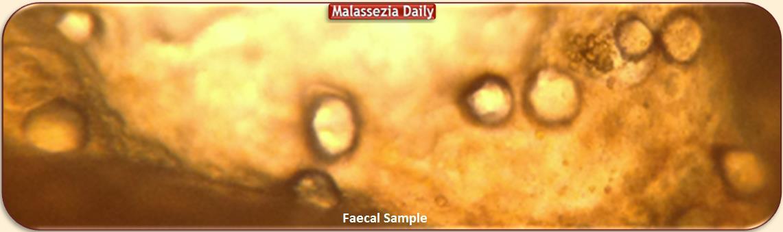
Current Faecal Samples
Another Surprise!
I guess there is no need to point out that
* I am No Professional Microbiologist
* Faecal material is Rich in Elements
i could not even imagine existing
* There could be many things staring me in the eye
i would have absolutely no clue
that they are staring me in the eye or what they are…
So!… Not having pointed this out…
I shall Point out that:
Here I am Only Looking for Malassezia Yeast / Fungus
in its Different Stages of Development
the way i have come to Recognise them
and it is on this basis
i will be presenting the Photos of the samples below
-This including possibilities of Error.
(No Microscope Oil has been used for any of these Samples)
Faecal Sample 1
View of Part 1 of Sample 1
Photo 1 and 2 Show a number of Large Circular Shapes
looking rather too large to be Yeast
compared with the size viewed in other samples
and quite Irregular to be Fluid Reflection of the Microscope’s lenses

Zoomed in
I called My Man – even more of an amateur than i am – for a
‘What do you think you See’
and he suggested they looked like ‘Fat globules’
-clever man!- and somehow i agree… but what kind of fat? …
‘Fat’ as Faecal matter Fat or as Malassezia ‘Fatty’ globule / blobs?
Certainly a good chapter of confusion and uncertainty this one 😦 …
Dec 14
Editing this Entry i Noticed 2 things:
1- The “Fat’ globules have a slight hint of a protrusion…
that normal Fat globules do not have
2- Perhaps the Darker outer Peripheries
are the Developing Familiar Semi – Circular
Beaded/Spore Eyelets ???
If that is so, it would explain why they are -initially larger in size
as being in earlier stage of development
and why i could not quite identify them with the same confidence.
View of Part 2 of Sample 1
Photo 1 Shows more of the same circular shapes
Photo 2 Shows a stray dead Malassezia hyphen

Faecal Sample 2
View of Part 1 of Sample 2
The same Circular Shapes

Below comparison with Google Malassezia Images

Looking at the Photos above and comparing with my samples
something tells me ‘yes, similar … perhaps identical
…yet … not Quite…’ but i cannot tell why not…
Perhaps because all spherical shapes i have seen in previous
faecal samples were much smaller without the ‘Fatty’ full look
and usually scattered around very long ‘nodulated’ hyphae
much like the second Google Photo above.
(See Note Above)
Comparing My Samples with Google Photos
There may be Difference in Microscopes
or Method, i.e. use of Oil, Focusing and /or Magnification
that may make them look similar or different…
I usually prefer the ‘almost identical’!
Comparing My Previous with the Current Samples
The Microscope is the same, and so is the Magnification.
Only the Focusing is adjusted where different parts
of the sample come clearer or go out out of focus
so Perhaps -and more likely to my guessing judgement-
the difference may be in Different Stage of Development?
and /or Yeast versus Fungus…
Part 2 of the Sample 2
The same Circular Shapes but also presence of Blood in two areas
making the ‘Malassezia Fatty Globules’ theory much more likely

But there is More!
I left the sample on the slide until the next day
to see if there would any detectable changes.
Part 2 of the Sample 2
Viewed the Next Day
The ‘Fatty globules’ seem to have Changed overnight

They look fuller and augmented … much like yeast does…
Some have augmented so much that they Bump onto Each Other
as shown Below
Day One Day Two

Changing Microscope Focusing deliberately but on that Same Spot
the Circular shapes go out of focus but Something Else comes to View

( Click on photo to enlarge )
Right below the Circled Big Blood Blob
looks like another dangling empty, dead Malassezia Hyphen
Below Zoomed in

So, to Sum it Up:
1) Spherical Shapes that seem to Augment
2) Blood Presence
3) Two Empty, Dead Malassezia Hyphae
All Pointing to Malassezia at the Yeast stage.
* * *
Below Comparing the Original Photo
taken with a Poor quality iPhone camera
that could pick up a plenitude of ‘Hyphae ‘Spaghetti’
but not the Spherical ‘Meatballs’ scattered around them

With the Current Sample Findings
of
1) A Number of Yeast Cells?
2) Blood Presence i.e Cloning stage of Development
3) Only Two Isolated empty Hyphae

and Considering that the Original poor quality Photos
did not present accurately all of what was Visible
I would say there is a marked Improvement
even in just that there are Not as many Yeast Cells
compared to Spaghetti Filaments as well as Scattered Cells
especially if most current Cells never reach maturity to Cloning Stage
which -IF this would be the case- would be due to
the Acidophilus Treatment
and Frequent Clearing of the Colon with Enemas
* * *
(NEXT: ‘ Malassezia – Saliva and Lung Blood Samples’ )

