To View Part 1 or 10 First
Click on Links Below
Malassezia: What The Photos Reveal – (PT 1)
Malassezia: What The Photos Reveal – (PT10)
*
Malassezia: What The Photos Reveal – (PT 11)
Malassezia
Reflection – Saliva and Lung Blood Samples
A Last Minute Temptation!
Clearing Unsuitable for Use Photos from my Folders
i caught a perfect one of a Reflection of the Microscope Lens
onto the sample

A few days back on the same day i collected the Vagina Sample
i had also placed a Saliva sample on a slide.
The First View of Both was absolutely mystifying for a few seconds

until the penny dropped that every Saliva Bubble
was reflecting the Microscope Lens back
A few hours later when most Bubbles had dried out
i re-adjusted slightly the focusing and some flattish circles
came to view that i could not easily identify
so i discarded the sample and concentrated on the Vaginal one.
All the while, thinking that if i had found
Fluorescent Green Malassezia in Lung Sputum
it was only Logical there Must Be present in the Saliva as well
especially since it seems to gather around my lips
practically after every time i have eaten my Fruities.
So when i got a sudden Lung Bleed it presented a last minute
temptation and opportunity to find out:
a) What i could see in the Lung Blood Sample, and
b) If Any Malassezia Presence in Blood or Surrounding Saliva
Here are the Results:
The First View of the Lung Blood was pretty much consistent
with the Microscope Reflection so i thought it was a lost case
and did not bother for all the trouble and effort it takes to try
to align the camera over the Microscoper Viewer to take a Photo
but i left the sample on the slide for ‘just in case’ there was
something worth viewing after drying up… and i was not wrong.
Lung Blood – Spot 1 – Second View 6-8 hours later

onsistent with the Previous Samples in the series
*Malassezia: What The Photos Reveal*
Obvious Malassezia Budding – if the Identification has been correct.
Lung Blood – Spot 1 – Next Day
No Further Changes detected

Lung Blood – Spot 2 – Viewed also the Next Day

Day 3
That Intuitive urge i get when i tune into things whispered again:
‘Check one more time’
I expected it would all be dead and dry by now
since there had been No change on Day 2’
but since i had to retrieve and clean the slide
i thought i might as well have another look.
And the ‘Whisper was Right!’…
A Last minute look
Perhaps Finally Brought the Proof needed!
A Budding Yeast with a Cloning Collaret???

Still No Hyphae anywhere big or small
but this looks pretty much like
a Cloning Collaret Budding Yeast!
Perhaps the Proof needed?…
Malassezia Globosa?…

Comparison with Grit in Eye Liquid — and Google Malassezia Images
1) Lung Blood – 2) Grit in Eye – 3) Google Malassezia Images
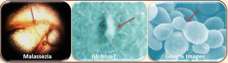
(Click on Images to Enlarge)
Comparison with Malassezia Globosa Google Images

My Verdict
Malassezia Everywhere – Inside Out
Contained and Controlled
as Much as Can Possibly Be Externally
but generally Inaccessible Internally.
I do Not give up easily so i intend to trial an idea
i have been considering for quite sometime
on how to administer Acidophilus Bacteria
as Close and Directly to the Lung as Possible
Bypassing Stomach Acids and Enzyme interference
The Official Stance…

Hard to tell which one, whether Furfur or Globosa
or any of the 11 Species Identified
(PART 12: * 1)The Colour Kaleidoscope – 2)The Common Characteristics *)

