* UNDER THE MICROSCOPE *
*

*Malassezia ~ New Discoveries and Photos Confirming Old (PT2) *
*
* Malassezia~Acidophilus and the Creamy Yellow Blobs *
PART 2
The day after the Lucky discovery of the
“Acidophilus and the Fluorescent Green”
Ears still treated with the Friendly –to humans– Bacteria
i used a Cotton tip to clean them and re-apply fresh dosage.
And here comes Surprise number 2!
After more than two years, if i recall correctly,
of never having collected anything ‘visible to the naked eye’
from the inside area of the ears, here come
not just one but two shapes of an old Familiar
‘worm looking like creature’ of a long time back,
just like the one below posted in the PHOTOS Section
on * Malassezia Blog *

Both of the New ones were the exact shape like the old one above.
My Phone camera could not ‘see them’ though, so i could not take a photo.
Not deterred by the set back and determined to ‘catch them’ while i could
i immediately unpacked Both Microscopes and tried the Digital one first
as it is the one that could take a magnified photo of the Cotton Bud
without disturbing the Blobs – just like the above .
Unfortunately i stumbled on a second setback:
My Laptop would not recognise the Digital M even though it was installed.
I had no idea where to find the CD to re-install it and no patience
to wait till dark when my man –keeper of Software CD’s– came home
so i proceeded to plan B, i.e try viewing under the Biological Microscope!
One problem with that was that it cannot ‘see’ through the cotton Bud
so i had to scrape the Blobs as carefully as possible and transfer them
onto the slide… and in my excitement did not think to scrape only one…
so both Blobs were a bit squeezed and lost their characteristic shape
– the reader will have to take my word that both blobs
looked like twins of the above old photo–
but despite this additional setback there were a couple more revelations!
Well after the Transfer it just Finally dawned on my consciousness
a bit too late to avoid the ‘disfigurement’ of the Blobs…
that for the past full year i had no reason to rely any more
on my poor quality phone camera
since my man had bought me for my Birthday
a beautiful Apple iPad with an Excellent camera!
So i tried it immediately to check how well it could ‘see’ them
directly on the slide – but not through the BM*
Here is how they ended up looking on the slide
after been gently scraped off the Cotton Tip

They Both have retained a slight semblance
of their initial elongated wormy shape
Then i had a great deal of trouble aligning
and focusing the i Pad Camera Eye
–being on the top left edge of its rectangle shape-
with the Cylindrical Single Top Eye of the Bio- Microscope
but the end result after several ‘hit and miss’ tries
was much better than any other photos i have presented before.
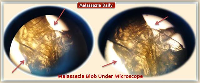
Below we can see what the Blobs looked under the Bio–Microscope.

The Elongated shape of this Blob is undeniable here
as well as its Yellow Creamy Colour and consistency.
And partly of the other Blob below
These two Blobs were collected 20 minutes after having applied
Acidophilus treatment and it was interesting that viewed with naked eye
and photographed with camera they look like Creamy wormy Blobs
but viewed under the Bio-Microscope they somewhat retain some
of the above characteristics and look more like the hyphae type of shards
as the older photos shown below
Original Photos – taken long time Back

and the two New ones together

but without any hyphae
perhaps caught at an earlier stage either not mature enough or dead.
Looking more closely though there are some distinctly visible
Long Filament like lines (hyphae?)

… just about ready to separate from the blobby gel
and spring outwards seeking Blood?
(Easy to see a resemblance of a Dr Who extraterrestrial style
of ‘Alien life’
and a possible alignment with what is called …Morgellons?
But Morgellons it is Not!… )
A Pre Solidified Shard with at least two visible Hyphae:
One or possibly two already impregnated with Blood
which – if proves right – adds another significant detail
to my theory during previous observations,
that it digs in -after it solidifies- to get blood
in that perhaps it can also do so both before as well as after.
For this purpose i have deliberately left the samples on the slide
in order to go back and check if there is any development
during the next few days.
The million dollar question is:
“Would they be able to develop further
after been treated with Acidophilus Bacteria?”
I have a photo, i will publish in a following Part of this topic
which casts doubt that it will be able to do so
unless it had already done so prior to Acidophilus
– as might be indicated above.
(DM* Digital Microscope)
(BM* Biological Microscope)
* * *
For your Convenience:
Malassezia ~ New Discoveries–Photos–Confirmations(1)
Malassezia ~ New Discoveries–Photos–Confirmations(2)
Malassezia ~ New Discoveries–Photos–Confirmations(3)
Malassezia ~ New Discoveries–Photos–Confirmations(4)
Malassezia ~ New Discoveries–Photos–Confirmations(5)
Malassezia ~ New Discoveries–Photos–Confirmations(6)
Malassezia ~ New Discoveries–Photos–Confirmations(7)
(Part 8 to Follow)
==============================================
Recommended Reading Entries in Order of Posting
* MALASSEZIA BLOGS *
![]()
* Malassezia Blog *
Photos – History of Treatments and Results
![]()
* Malassezia Daily *
New Photos – New Discoveries
Treatments and Results
![]()
Malassezia Issues – Related Topics
Readers Questions Answered
Malassezia and Medical Research
Health and Immunity – Nutrition Diet
=============================================


12 responses to “Malassezia~New Discoveries-PhotosConfirmations(2)”